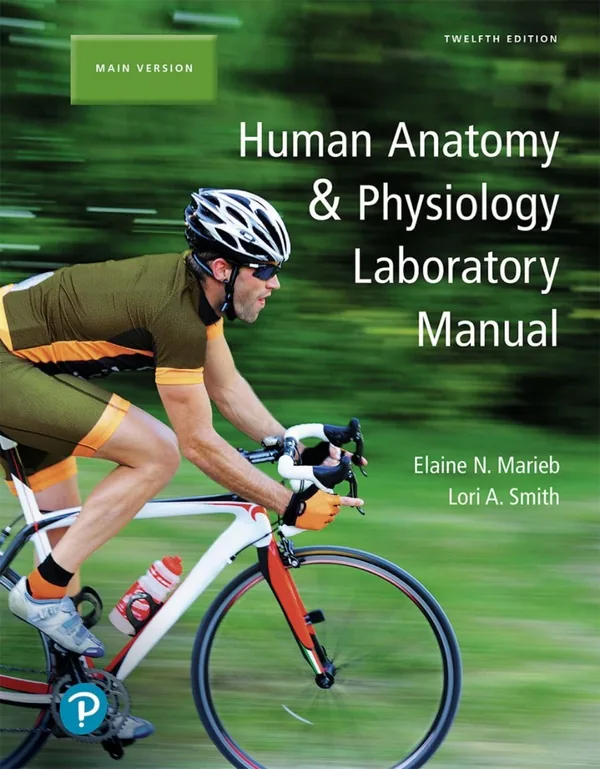
Human Anatomy & Physiology Laboratory Manual, 13th Edition, provides laboratory exercises for undergraduate study of the human body. Elaine N. Marieb and Lori A. Smith move from terminology, cells, and tissues to organ-system anatomy and physiological measurements. Illustrations and clinical application questions connect laboratory observations with concepts used in health-science courses.
Inside this title
Anatomical terminology, body organization, cells, and tissue identification
Current listed price $19.00Regular price $38.00Digital ebook
Designed around a two-semester anatomy and physiology laboratory sequence, this manual combines structural identification with experiments exploring function. Early work addresses anatomical directions, body organization, cell transport, mitosis, and tissue recognition. Skeletal and muscular exercises then develop familiarity with bones, joints, and muscle actions.
System-based activities include nervous-system anatomy and reflexes, heart examination, blood-pressure measurement, respiratory testing, and study of digestive, urinary, and reproductive structures. Figures and photographs support observation, while clinical application questions invite students to interpret what their findings mean. Nursing and allied health students, other undergraduate learners, and laboratory instructors can use the exercises to organize practical study alongside an anatomy and physiology course.
Study guide included
Inside Human Anatomy & Physiology Laboratory Manual, 13th Edition.
This manual teaches by doing: students identify structures, run comparisons, and record measurements rather than only reading descriptions. Each exercise links something seen on a model or slide to a function tested in the lab, and review sheets ask learners to explain results instead of just reporting them.
First exercises cover anatomical position, regional terms, planes, cavities, and microscopy, including field diameter and wet mounts. Later work moves through cell structure, membrane transport, histology, bones, joints, muscle, nervous tissue, reflexes, and sensation. Blood, heart and vessels, ECG and pulse, respiration, digestion, urine testing, reproduction, heredity, and PhysioEx simulations follow. The routine is consistent: prepare beforehand, document carefully, and use review sheets to interpret what happened. Instructors may follow the sequence or pick exercises that fit a term. Safety and care with specimens matter.
Orientation and trustworthy lab observation
Anatomical position anchors directional vocabulary, while regional terms, planes, and cavities place what a specimen displays. A part seen from an odd angle or cut in section may look strange, so record the viewpoint and adjacent landmarks before transferring a label. Microscopy adds scale: power alters the visible field and what remains sharp, and a sketch should carry its view, magnification, and edges.
Fix the view before naming a part
Higher power shrinks the visible field
Sketch scale so others can relocate it
Landmarks anchor a transfer from diagram
Body orientation and microscopy exercises
Cells, tissues, and movement experiments
Models stress membranes, nuclei, and organelles; real specimens show how much those features differ. Recognizing tissue requires attention to arrangement, not only individual cell outline: epithelia present surfaces and layering, connective tissue shows abundant matrix, and muscle or nerve carries specializations suited to its role. Transport labs distinguish diffusion, osmosis, and filtration by varying conditions and asking which process accounts for the observed direction.
Arrangement matters as much as cell shape
Matrix dominates in connective tissue
Dye movement shows a concentration gradient
Applied pressure can push fluid through a filter
Cell anatomy, membrane transport, and histology exercises
Bones, joints, and muscle traces
Long-bone study ties fine structure to visible layout, while exercises on skull, vertebrae, girdle, and limbs situate landmarks within the axial versus appendicular divisions. Joint labs show how surfaces and stabilizing tissues limit motion. Muscle histology and neuromuscular junction work set up contraction recordings, where stimulus timing and recording conditions explain how a force curve evolves and tires.
Describe the joint before naming a muscle
Growth regions show up in long bone
Compare traces on the same timescale
Fatigue lowers output across repeated work
Bone, joints, skeletal muscle, and muscle physiology exercises
Nerves, reflexes, and sensory thresholds
Students map brain regions, cranial nerves, spinal structures, and peripheral plexuses, then use reflex activities to link a sensory input with an output along a defined route. Sensation labs cover touch, adaptation, vision, balance, hearing, smell, and taste. A two-point threshold reflects receptor density and testing discipline, so matching contact, spacing, and instructions makes regional comparison meaningful.
Reflex arc links input to output
Pupil change gives visible evidence
Threshold tracks how densely receptors sit
Repeat trials beat one odd response
Brain, spinal cord, reflexes, and sensory exercises
Signals, gases, and filtrate measurements
Blood labs divide cells from plasma and introduce measures such as hematocrit. Models of the heart and vessels trace pulmonary and systemic paths. An ECG captures electrical activity; heart sounds and a felt pulse mark mechanical events, and comparing their timing links several views of one cycle. Exercises on respiration, digestion, and urine each ask students to identify what was measured and what bodily process it reflects.
Electrical events differ from mechanical ones
Align signals before comparing timing
Name the variable and its process
Enzyme action depends on conditions
Blood, cardiovascular, respiratory, digestive, and urinary exercises
Development, heredity, and review
Slides and models allow comparison of reproductive organs, gamete formation, embryos, the placenta, and fetal structures. Meiosis alters chromosome count and assortment, and staging a section means checking which structures appear and where the cut passes. Heredity work uses crosses and probability to state expectations, while blood groups and electrophoresis show how an observed phenotype can rule out some genotypes.
A single slide shows only one moment
Meiosis reshuffles and halves chromosomes
Predict ratios before checking a sample
Review sheets expose gaps for a second pass
Reproduction, development, heredity, and PhysioEx exercises
Chapter and section map
Locating Body Regionsp. 3
Preparing and Observing a Wet Mountp. 31
Demonstrating Movements of Synovial Jointsp. 170
Studying the Microscopic Structure of Selected Neuronsp. 256
Tracing the Path of Blood Through the Heartp. 439
Studying Nephron Structurep. 608
Exploring Probabilityp. 669
PhysioEx Skeletal Muscle PhysiologyPEx-17
Key takeaways
State orientation, scale, and a visible landmark before you identify any structure.
Hold one comparison condition steady and document your method before explaining a difference.
Explain how a signal maps to a process without treating the two as identical.
Undergraduate anatomy and physiology lab students in nursing, allied health, and biology programs, plus instructors seeking structured exercises with review and simulation components for a full term.
Topics covered
anatomy lab manual
microscopy exercises
membrane transport
tissue histology
skeletal muscle physiology
reflex testing
PhysioEx simulations
urine analysis
Questions about this ebook.
Edition, audience, and study-guide details at a glance.
What is Human Anatomy & Physiology Laboratory Manual, 13th Edition about?
Human Anatomy & Physiology Laboratory Manual, 13th Edition, provides laboratory exercises for undergraduate study of the human body. Elaine N. Marieb and Lori A. Smith move from terminology, cells, and tissues to organ-system anatomy and physiological measurements. Illustrations and clinical application questions connect laboratory observations with concepts used in health-science courses.
Which edition of Human Anatomy & Physiology Laboratory Manual, 13th Edition is this?
This listing is for the 13th edition. Check the title, ISBN, and book details above before ordering, especially if your course or reading list names a specific edition.
Who is Human Anatomy & Physiology Laboratory Manual, 13th Edition for?
Undergraduate anatomy and physiology lab students in nursing, allied health, and biology programs, plus instructors seeking structured exercises with review and simulation components for a full term.
Does this page include a study guide?
Yes. This page includes an original study guide with six thematic sections, key takeaways, and a chapter and section map for structured reading.