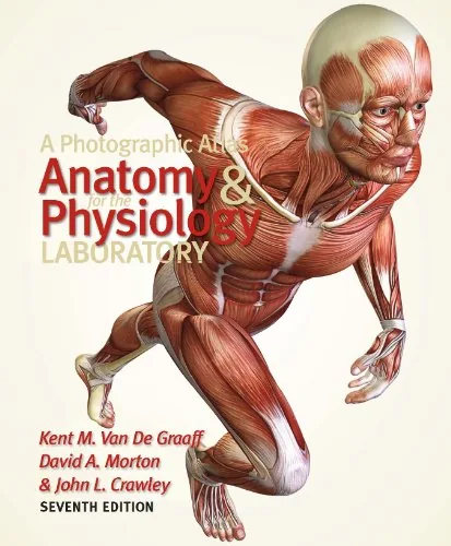
A Photographic Atlas for the Anatomy and Physiology Laboratory, 7th Edition
By Kent M. Van De Graaff, David A. Morton, John L. Crawley
About this ebook
A Photographic Atlas for the Anatomy and Physiology Laboratory, Seventh Edition supports visual study of human anatomy through labeled photographs. Intended for anatomy, physiology, nursing, and allied health students, it presents skeletal, muscular, nervous, cardiovascular, and other organ systems, helping readers relate anatomical terminology to structures and their spatial relationships.
Inside this title
Labeled anatomical photographs emphasize real tissue and spatial relationships
Current listed price $14.99Regular price $42.00Digital ebook
Photographs of anatomical specimens provide the central teaching material in this laboratory atlas. Labels and leader lines identify structures, allowing students to compare the appearance of real tissues with the terminology introduced in lectures and textbooks. The photographic approach emphasizes relationships that may be less apparent in simplified drawings.
Coverage extends across bones and joints, muscles, the heart and vessels, respiratory and digestive organs, the nervous system, and urinary and reproductive structures. The seventh edition is intended to accompany anatomy or integrated anatomy and physiology courses. Students preparing for laboratory identification work and instructors seeking visual teaching material can consult the body-system organization alongside their course's laboratory activities.
Study guide included
Inside A Photographic Atlas for the Anatomy and Physiology Laboratory, 7th Edition.
Anatomy is learned twice: once as vocabulary and once as the look of real tissue. This photographic atlas guide follows that second learning, moving system by system through labeled specimens so that terms, positions, and neighbouring structures can be studied together in preparation for laboratory identification work.
Photographs occupy the center of this laboratory atlas because dissection and identification depend on recognizing actual tissue. Each system is presented through labeled images, with leader lines tying terms to structures and their neighbours. The organization follows body systems: bones and joints, muscles, the nervous system, the heart and vessels, respiratory and digestive organs, and the urinary and reproductive structures. Because the images retain the texture and spatial relations of specimens, students can rehearse what they will see in the lab. The atlas is a companion for courses and practical identification work, not a substitute for either.
Learning anatomy from photographs
Photographs preserve the appearance of specimens that drawings simplify away. The atlas uses that fidelity to teach recognition of tissue, colour, and spatial relation, including structures that are easy to miss in a diagram. Students are asked to match terminology to what they actually see.
Photographs retain texture and spatial relation
Labels connect terms to visible structures
Recognition supplements what diagrams show
Topics 1, 8
Bones and joints
The skeletal material identifies individual bones and the features that mark them, then places them in relation to neighbours and joints. Learning the landmarks is presented as a way of reasoning about position, which matters when a structure must be located on a specimen rather than named in isolation.
Landmarks mark individual bones
Joints place bones in relation to one another
Position matters for laboratory identification
Topic 2
The muscular system
Muscle coverage connects each muscle with its attachments and its place among surrounding structures. Because photographs show actual fibres and fascial relationships, the student works with form rather than a schematic outline. The goal is recognition that transfers to laboratory identification.
Muscles are tied to attachments
Fascial relationships are visible in specimens
Form supports recognition in the lab
Topic 3
The nervous system
The nervous system is treated through its visible organization, from central structures to peripheral arrangements. The atlas uses labeled images to make regions and pathways identifiable, keeping the terminology attached to something observable. This supports study of a system that is otherwise difficult to visualize.
Central and peripheral organization is shown
Labels keep terms tied to observable regions
Visual study aids a hard-to-see system
Topic 4
Heart, vessels, and breathing
Cardiovascular and respiratory material shows the heart, vessels, and airway structures as they appear in specimens. The photographs emphasize continuity and adjacency, so that chambers, great vessels, and respiratory organs are understood in relation to one another rather than as separate lists.
Chambers and vessels are shown in continuity
Adjacency links organ systems
Respiratory structures are studied in place
Topics 5-6
Digestion, excretion, and reproduction
Digestive, urinary, and reproductive organs are presented with attention to position and neighbouring structures. The images give students a way to study organs that are often represented only schematically, and to connect gross appearance with the terminology used in lecture and laboratory.
Organs are studied with their neighbours
Photographs counter oversimplified diagrams
Appearance is linked to terminology
Topics 6-7
Topic map
Reading labeled anatomical photographsTopic 1
Skeletal system and jointsTopic 2
Muscular systemTopic 3
Nervous systemTopic 4
Cardiovascular systemTopic 5
Respiratory and digestive systemsTopic 6
Urinary and reproductive systemsTopic 7
Terminology and spatial relationshipsTopic 8
Key takeaways
Photographs teach recognition that diagrams alone cannot provide.
System organization helps students relate structures rather than memorize lists.
The atlas works best alongside laboratory activity and lecture terminology.
Anatomy and physiology students, nursing and allied health learners, and instructors who need visual material for laboratory identification work.
Topics covered
photographic anatomy atlas
anatomy and physiology laboratory
labeled anatomy photographs
skeletal system
muscular system
nervous system
organ systems
allied health anatomy
Questions about this ebook.
Edition, audience, and study-guide details at a glance.
What is A Photographic Atlas for the Anatomy and Physiology Laboratory, 7th Edition about?
A Photographic Atlas for the Anatomy and Physiology Laboratory, Seventh Edition supports visual study of human anatomy through labeled photographs. Intended for anatomy, physiology, nursing, and allied health students, it presents skeletal, muscular, nervous, cardiovascular, and other organ systems, helping readers relate anatomical terminology to structures and their spatial relationships.
Which edition of A Photographic Atlas for the Anatomy and Physiology Laboratory, 7th Edition is this?
This listing is for the 7th edition. Check the title, ISBN, and book details above before ordering, especially if your course or reading list names a specific edition.
Who is A Photographic Atlas for the Anatomy and Physiology Laboratory, 7th Edition for?
Anatomy and physiology students, nursing and allied health learners, and instructors who need visual material for laboratory identification work.
Does this page include a study guide?
Yes. This page includes an original study guide with six thematic sections, key takeaways, and a topic map for structured reading.