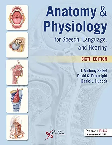
Anatomy & Physiology for Speech, Language, and Hearing, 6th Edition
By J. Anthony Seikel, David G. Drumright, Daniel J. Hudock
About this ebook
Anatomy & Physiology for Speech, Language, and Hearing, 6th Edition examines the body systems that support communication. Seikel, Drumright, and Hudock connect respiratory, phonatory, articulatory, auditory, and neural function. Illustrations and clinical examples support students in speech-language pathology and audiology studying normal communication and the anatomical basis of disorders.
Inside this title
Respiratory mechanics, laryngeal structures, and vocal fold function
Current listed price $26.00Regular price $58.00Digital ebook
Speech and hearing depend on coordinated activity across several body systems. This text follows breathing and pressure generation into the mechanics of the larynx, vocal fold vibration, and the structures that shape speech sounds. Peripheral and central auditory pathways extend the account from sound reception to neural processing.
Anatomical illustrations accompany explanations of function and clinical examples involving dysarthria, aphasia, and hearing loss. The sixth edition also considers cranial nerves, cortical centers, and developmental changes from infancy to adulthood. Students of speech-language pathology and audiology can use its system-by-system coverage to connect anatomical terminology with normal and disordered communication.
Study guide included
Inside Anatomy & Physiology for Speech, Language, and Hearing, 6th Edition.
A focused account of the respiratory, phonatory, articulatory, auditory and neural systems behind speech, voice, hearing and swallowing. Seikel, Drumright and Hudock write for speech-language pathology and audiology students who need anatomy to explain how communication works and where it can break down.
After orientation and tissue basics, paired chapters treat breathing, phonation, articulation and resonance, with a separate chapter on chewing and swallowing. The final chapters follow sound from the outer ear through cochlear transduction and then trace the neural pathways that support perception and motor control. Illustrations, clinical connections and study questions keep detailed anatomy attached to observable communication, so readers can reason from structure to function rather than memorizing labels. Each chapter pairs a structural account with the function it enables.
Orientation, planes and shared structures
Planes and directional terms give one reference for describing the body, and movement terms state what a structure does relative to a joint or landmark. Because small positional shifts alter airflow or signal paths in speech and hearing, the starting position and direction must be named. Tissues range from lining epithelium to force-producing muscle and signal-carrying nerve, and one structure, such as the pharynx, can serve airflow, resonance and swallowing.
Planes and direction terms anchor descriptions
Small shifts can change airflow or signal paths
Tissues differ in properties and roles
Shared structures serve several functions
Ch. 1 · Basic Elements of Anatomy
Respiration for speech
Thoracic structures expand and contract, and during inspiration the diaphragm and other muscles change volume so pressure differences draw air in. Quiet expiration leans heavily on elastic recoil, while lung volumes, posture and airway resistance alter the resulting flow. Speech takes air in quickly and releases it slowly, with respiratory muscles checking or assisting recoil to supply pressure and flow suited to phonation and articulation.
Volume changes create pressure and airflow
Elastic recoil drives quiet expiration
Speech uses quick intake and slow release
Breath groups reflect phrasing and air supply
Chs. 2–3 · Anatomy and Physiology of Respiration
Phonation and vocal control
Laryngeal cartilages and intrinsic muscles open, close and adjust the folds for breathing, voicing and protection. Once positioned with adequate pressure, tissue elasticity and aerodynamic forces sustain vibration, and the text separates onset, maintenance and offset. Vibration frequency contributes to pitch through length, tension and vibrating mass, while pressure and fold behavior change source intensity, so a listener's impression alone cannot identify the adjustment.
Intrinsic muscles position the vocal folds
Elasticity and airflow sustain vibration
Length and tension shift pitch
Loudness and pitch can interact
Chs. 4–5 · Anatomy and Physiology of Phonation
Articulation and resonance
The jaw, lips and tongue reshape the oral cavity and form constrictions, while the hard palate and teeth provide landmarks and the velum couples or uncouples the nasal cavity. The source-filter model separates the laryngeal source from vocal-tract shaping, which explains why one source yields different vowel qualities. Real articulation is continuous, with neighboring sounds influencing position, and sensory feedback links intended and produced movement.
Articulators reshape the resonating cavities
The velum controls nasal coupling
Source-filter separates voice from tract shape
Coarticulation links neighboring sounds
Chs. 6–7 · Articulation and Resonation
Chewing and swallowing
Mastication combines jaw movement, tongue positioning and saliva to prepare a bolus, and oral transport carries it toward the pharynx while sensation reports what the material is and where it sits. Pharyngeal events move the bolus through a region shared with the airway, with timed muscular and protective actions directing it toward the esophagus. Sensory input, voluntary action and reflex patterns interact, so a single observation cannot identify the phase at fault.
Chewing prepares material for transport
Sensation reports bolus position
Pharyngeal timing protects the airway
Reflex and voluntary control interact
Ch. 8 · Physiology of Mastication and Deglutition
Hearing and neural coordination
Sound passes through the outer ear to the tympanic membrane and middle-ear ossicles, and movement at the cochlea displaces fluid and membranes. Regions of the basilar membrane respond best to particular frequencies, giving the system an organized starting point. Hair cells convert that movement into electrical change, auditory pathways ascend through brainstem and higher centers, and vestibular structures report head motion, while neural pathways coordinate sensory and motor function.
Middle-ear ossicles transmit mechanical energy
Basilar membrane regions tune to frequency
Hair cells transduce movement into signal
Vestibular input reports head orientation
Chs. 9–12 · Hearing, Auditory Physiology and Nervous Systems
Chapter and section map
Basic Elements of AnatomyCh. 1 · p. 1
Anatomy and Physiology of RespirationChs. 2–3 · pp. 47, 145
Anatomy and Physiology of PhonationChs. 4–5 · pp. 185, 247
Articulation and ResonationChs. 6–7 · pp. 305, 417
Mastication and DeglutitionCh. 8 · p. 455
Anatomy and Physiology of HearingChs. 9–10 · pp. 515, 549
Neuroanatomy and NeurophysiologyChs. 11–12 · pp. 601, 745
Key takeaways
Describe a structure by its location, the movements it makes and the task it supports.
Separate respiratory power, the laryngeal source and vocal-tract filtering when explaining speech.
Follow sound through mechanical transmission, cochlear transduction and neural processing as distinct stages.
Students in speech-language pathology and audiology programs, and instructors teaching a dedicated anatomy and physiology course for communication sciences and disorders and its clinical foundations.
Topics covered
speech anatomy
respiration for speech
laryngeal phonation
vocal tract resonance
mastication and deglutition
auditory physiology
cranial nerves
communication sciences
Questions about this ebook.
Edition, audience, and study-guide details at a glance.
What is Anatomy & Physiology for Speech, Language, and Hearing, 6th Edition about?
Anatomy & Physiology for Speech, Language, and Hearing, 6th Edition examines the body systems that support communication. Seikel, Drumright, and Hudock connect respiratory, phonatory, articulatory, auditory, and neural function. Illustrations and clinical examples support students in speech-language pathology and audiology studying normal communication and the anatomical basis of disorders.
Which edition of Anatomy & Physiology for Speech, Language, and Hearing, 6th Edition is this?
This listing is for the 6th edition. Check the title, ISBN, and book details above before ordering, especially if your course or reading list names a specific edition.
Who is Anatomy & Physiology for Speech, Language, and Hearing, 6th Edition for?
Students in speech-language pathology and audiology programs, and instructors teaching a dedicated anatomy and physiology course for communication sciences and disorders and its clinical foundations.
Does this page include a study guide?
Yes. This page includes an original study guide with six thematic sections, key takeaways, and a chapter and section map for structured reading.