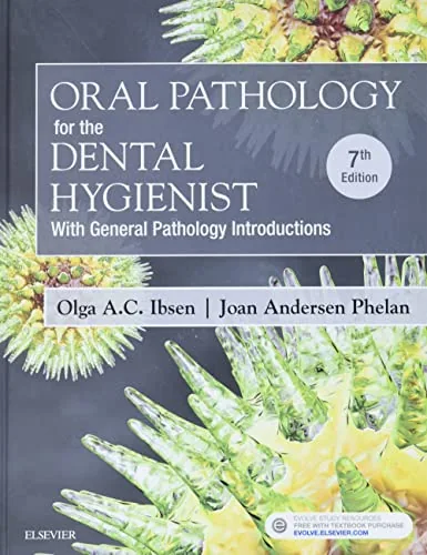
Oral Pathology for the Dental Hygienist, 7th Edition
By Olga A.C. Ibsen, Joan Andersen Phelan
About this ebook
Oral Pathology for the Dental Hygienist, seventh edition, relates general pathology to oral diseases and lesion recognition. Ibsen and Phelan introduce inflammation, repair, and neoplasia before examining conditions affecting oral tissues. Dental hygiene students can study clinical and radiographic findings, systemic disease manifestations, and the hygienist's role in assessment and education.
Inside this title
General pathology introductions explaining inflammation, repair, and neoplasia
Current listed price $32.99Regular price $85.00Digital ebook
General pathology introductions establish the processes behind tissue injury, inflammation, healing, and abnormal growth. The oral disease coverage then links these mechanisms to the appearance, causes, and clinical significance of soft and hard tissue lesions. Comparisons of clinical and radiographic features provide a basis for studying differential diagnosis.
Topics include oral manifestations of systemic disease, fungal and viral infections, inherited conditions, and oral cancer risk and recognition. The seventh edition also discusses HPV-related oropharyngeal cancers and autoimmune disorders. Its dental hygiene focus keeps the relationship between pathology findings, the process of care, and patient education central to the material, making it suitable for students learning to connect classroom pathology with oral assessment.
Study guide included
Inside Oral Pathology for the Dental Hygienist, 7th Edition.
Ibsen and Phelan treat oral pathology as disciplined observation rather than pattern matching. The seventh edition pairs general pathology with lesion recognition, teaching hygienists to describe what they see, weigh clinical and radiographic evidence, and hand a clear preliminary record to the dentist. Its scope runs from normal variation through infection, developmental conditions, tumors and systemic signs.
The book starts with the diagnostic process and the limits of normal anatomy, then introduces injury, immunity and infection so that similar-looking lesions can be separated by mechanism. Developmental and genetic chapters explain how timing and distribution narrow a differential. Neoplasia and bone disease chapters organize tumors by tissue origin and teach readers to describe radiographic patterns before naming a condition. Later chapters place mucosal findings beside medical history, cancer therapy effects and orofacial pain. Throughout, synopsis tables are meant to be read with the patient record, not instead of it.
Describing before naming
The first chapter builds an evidence record across clinical, radiographic, historical, laboratory and microscopic categories. Precise description of site, border, color and texture lets another clinician follow the observation even when the cause is still open.
Record observable features in neutral terms
Compare against expected normal variation
Keep preliminary impression separate from diagnosis
Ch. 1, Introduction to Preliminary Diagnosis of Oral Lesions
Injury, inflammation and repair
Swelling, redness and ulceration signal that tissue is reacting, but they do not identify the trigger. The chapter links innate defenses and healing to traumatic and periapical processes, and it asks readers to locate a change anatomically before consulting comparison tables.
Separate the response from its cause
Anchor findings to the tissue of origin
Use history of trauma or irritation as context
Ch. 2, Inflammation and Repair
Immune and infectious mechanisms
Immune responses can protect or damage tissue, and infections arrive from bacterial, fungal and viral sources. Distribution, course and relevant history do the work that appearance alone cannot, especially where a patient's systemic status may be involved.
Distinguish defensive from damaging immune activity
Group pathogens and compare presentations
Treat a visible pattern as a starting clue
Chs. 3–4, Immunologic Oral Lesions and Infectious Diseases
Formation and inheritance
Developmental chapters follow the formation of face, oral cavity and teeth so that unusual shape or eruption has a reference point. Genetics supplies chromosome and gene background, while the text keeps a single oral finding from standing in for a whole diagnosis.
Link anomalies to the stage that produced them
Use imaging for changes inside bone
Scope genetic evidence to what it can support
Chs. 5–6, Developmental Disorders and Genetics
Tumors and bone patterns
Neoplasia is organized by tissue origin, with the caution that clinical appearance does not reliably separate benign, malignant and reactive growth. Bone chapters apply the same discipline to density, borders and effects on adjacent structures.
Sort tumors by tissue of origin
Describe an image before inferring a process
Expect overlap between categories
Chs. 7–8, Neoplasia and Nonneoplastic Diseases of Bone
Context beyond the lesion
Systemic illness, medication and cancer treatment can change what a mucosal finding means, and pain may be felt far from its source. The final chapters argue for recording patient reports and examiner observations as separate lines.
Read the medical history with the examination
Characterize pain by onset, duration and triggers
Note where symptoms are felt versus where they begin
Chs. 9–10, Systemic Diseases and Orofacial Pain
Chapter and section map
Preliminary diagnosis of oral lesionsCh. 1
Inflammation and repairCh. 2
Immunity and infectious diseasesChs. 3–4
Development and geneticsChs. 5–6
Neoplasia and bone disordersChs. 7–8
Systemic manifestations and orofacial painChs. 9–10
Key takeaways
Describe the lesion before choosing a category; description survives uncertainty.
Read comparison tables together with history and imaging, not as diagnosis.
State which evidence supports the impression and what still needs the dentist's assessment.
Dental hygiene students, practicing hygienists refreshing lesion recognition, and educators who teach the hygienist's role in oral assessment and patient education.
Topics covered
oral pathology
dental hygienist textbook
oral lesion recognition
clinical and radiographic findings
systemic disease oral signs
oral cancer recognition
dental hygiene education
Questions about this ebook.
Edition, audience, and study-guide details at a glance.
What is Oral Pathology for the Dental Hygienist, 7th Edition about?
Oral Pathology for the Dental Hygienist, seventh edition, relates general pathology to oral diseases and lesion recognition. Ibsen and Phelan introduce inflammation, repair, and neoplasia before examining conditions affecting oral tissues. Dental hygiene students can study clinical and radiographic findings, systemic disease manifestations, and the hygienist's role in assessment and education.
Which edition of Oral Pathology for the Dental Hygienist, 7th Edition is this?
This listing is for the 7th edition. Check the title, ISBN, and book details above before ordering, especially if your course or reading list names a specific edition.
Who is Oral Pathology for the Dental Hygienist, 7th Edition for?
Dental hygiene students, practicing hygienists refreshing lesion recognition, and educators who teach the hygienist's role in oral assessment and patient education.
Does this page include a study guide?
Yes. This page includes an original study guide with six thematic sections, key takeaways, and a chapter and section map for structured reading.