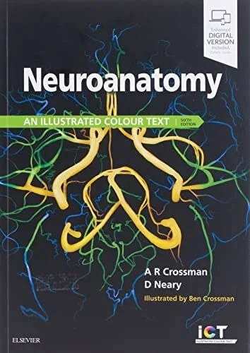
Neuroanatomy: An Illustrated Colour Text, 6th Edition
By Alan R. Crossman, David Neary
About this ebook
Neuroanatomy: An Illustrated Colour Text, Sixth Edition, introduces nervous system structure through color diagrams and clinical connections. Alan R. Crossman and David Neary write for health science students, covering sensory and motor pathways, cranial nerves, brain blood supply, cerebrospinal fluid, and the anatomical basis of neurological signs.
Inside this title
Color diagrams connecting nervous system structure and function
Current listed price $19.99Regular price $35.00Digital ebook
Visual explanation drives this introduction to the central and peripheral nervous systems. Diagrams accompany concise topic discussions so students can relate anatomical structures to their functions. Sensory and motor pathways are considered alongside the effects of lesions, bringing clinical interpretation into the study of anatomy.
Cranial nerve organization, vascular territories, the ventricular system, and cerebrospinal fluid circulation receive focused coverage. Neurotransmitter pathways and clinical examples extend the connection between structure, function, and disease presentations. The sixth edition's layout combines illustrations with clinical panels, making it useful for medical and other health science courses where students need to move between identifying a structure and understanding its functional significance.
Study guide included
Inside Neuroanatomy: An Illustrated Colour Text, 6th Edition.
Neuroanatomy: An Illustrated Colour Text introduces nervous system structure through diagrams and clinical correlations. Crossman and Neary guide health science students from sensory and motor pathways to cranial nerves, vascular supply, and cerebrospinal fluid, always linking anatomy to the neurological signs seen in practice.
The book treats neuroanatomy as a visual subject. Color diagrams sit beside concise explanations so students can identify a structure and immediately connect it to function. Pathways are traced from receptor to cortex, and lesion effects are described to show why the anatomy matters clinically. Separate sections cover cranial nerve organization, arterial territories, the ventricular system, and cerebrospinal fluid circulation. Neurotransmitter pathways extend the discussion toward function and disease. Clinical panels recur throughout, giving readers a way to test recognition against realistic presentations. The layout suits medical and health science courses that expect students to move between labeling a diagram and explaining a patient sign.
Structure and Function Through Diagrams
Illustrations carry much of the teaching load. Crossman and Neary pair each diagram with concise text so a structure, its connections, and its role can be learned together. Students are encouraged to read images as explanations, not merely as labels to memorize before an examination.
Read diagrams as explanations of function
Connect each structure to its connections
Use labels as part of a larger picture
Read at the start of each illustrated section
Tracing the Sensory Pathways
Sensory pathways are followed from peripheral receptors through relay stations to cortical areas. The book shows how a lesion at each stage alters sensation, turning a pathway diagram into a diagnostic tool. This progression supports clinical reasoning about sensory loss rather than isolated recall, and it gives students a way to organise examination findings.
Follow sensory routes from receptor to cortex
Locate relay stations along each pathway
Relate lesion sites to sensory deficits
Read with the pathway diagrams open
Tracing the Motor Pathways
Motor systems are presented with the same pathway logic, from cortical origin down to the relevant lower structures. Upper and lower motor patterns are distinguished through their effects. Students learn to predict weakness patterns from anatomical damage and to check those predictions against clinical examples.
Map motor routes from cortex downward
Distinguish upper and lower motor effects
Predict weakness patterns from lesion location
Read alongside lesion case panels
Cranial Nerves and Clinical Signs
Cranial nerve organization receives focused coverage, with attention to how each nerve's course explains its signs. The material links anatomy to examination findings so students can connect what they observe to underlying structure. Clinical examples reinforce the nerve-by-nerve detail and show how a single lesion can produce a recognizable pattern.
Learn cranial nerve organization and courses
Connect nerve anatomy to examination findings
Use clinical examples to reinforce detail
Read as a dedicated cranial nerve review
Blood Supply and Vascular Territories
Arterial supply is mapped by territory, making clear why particular vessel problems produce particular deficits. The book ties vascular anatomy to the functional regions each vessel serves. This supports interpretation of stroke patterns and ischemic presentations, and it helps students connect a clinical picture to a specific territory.
Map arterial territories of the brain
Relate vessel problems to specific deficits
Interpret stroke patterns through anatomy
Read with the vascular territory maps
Ventricles, CSF, and Neurotransmitters
The ventricular system and cerebrospinal fluid circulation are explained with attention to flow and obstruction. Neurotransmitter pathways add a functional layer beyond structure. Together these sections broaden the picture of how the nervous system maintains and modulates its activity, and they connect anatomy to conditions involving pressure or flow.
Trace cerebrospinal fluid flow and obstruction
Learn ventricular anatomy and relationships
Place neurotransmitter pathways within function
Read after the major pathway sections
Topic map
Nervous System OrganizationTopic 1
Sensory PathwaysTopic 2
Motor PathwaysTopic 3
Lesion CorrelationTopic 4
Cranial NervesTopic 5
Brain Blood SupplyTopic 6
Ventricular System and Cerebrospinal FluidTopic 7
Neurotransmitter PathwaysTopic 8
Key takeaways
Use each diagram as an explanation, tracing connections rather than memorizing isolated labels.
Practise predicting deficits from lesion sites; that habit converts anatomy into clinical reasoning.
Review cranial nerves and vascular territories together, since both explain common neurological presentations.
Medical and health science students taking neuroanatomy courses, plus learners who need a diagram-led revision text that links structures, pathways, lesion effects, and clinical signs.
Topics covered
neuroanatomy textbook
cranial nerves
sensory pathways
motor pathways
brain blood supply
cerebrospinal fluid
neurological lesions
illustrated colour text
Questions about this ebook.
Edition, audience, and study-guide details at a glance.
What is Neuroanatomy: An Illustrated Colour Text, 6th Edition about?
Neuroanatomy: An Illustrated Colour Text, Sixth Edition, introduces nervous system structure through color diagrams and clinical connections. Alan R. Crossman and David Neary write for health science students, covering sensory and motor pathways, cranial nerves, brain blood supply, cerebrospinal fluid, and the anatomical basis of neurological signs.
Which edition of Neuroanatomy: An Illustrated Colour Text, 6th Edition is this?
This listing is for the 6th edition. Check the title, ISBN, and book details above before ordering, especially if your course or reading list names a specific edition.
Who is Neuroanatomy: An Illustrated Colour Text, 6th Edition for?
Medical and health science students taking neuroanatomy courses, plus learners who need a diagram-led revision text that links structures, pathways, lesion effects, and clinical signs.
Does this page include a study guide?
Yes. This page includes an original study guide with six thematic sections, key takeaways, and a topic map for structured reading.