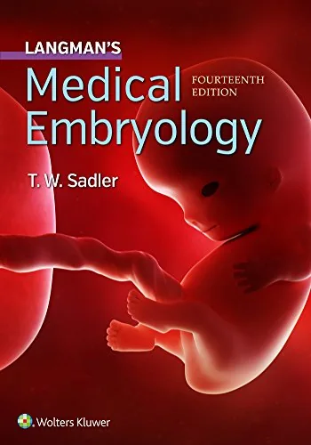
Langman's Medical Embryology explains human development through concise text, diagrams, and clinical correlations. T. W. Sadler's fourteenth edition moves from fertilization and early embryonic organization to the formation of organ systems, helping medical and health professions students relate developmental processes, molecular regulation, congenital anomalies, and the timing of harmful exposures.
Inside this title
Follows early embryonic stages through organ system development
Current listed price $9.99Regular price $16.00Digital ebook
The book divides embryology into early development and the development of individual organ systems. Fertilization, implantation, cleavage, gastrulation, and germ-layer formation establish the sequence before the cardiovascular, nervous, musculoskeletal, gastrointestinal, and urogenital systems are considered. Molecular and genetic regulation helps explain how these developmental events are coordinated.
Clinical correlates connect developmental mechanisms with congenital abnormalities, including neural tube defects, cleft palate, and septal defects. Illustrations and imaging examples support the spatial and chronological explanations, while chapter problems offer opportunities to apply concepts. The fourteenth edition is designed for medical and health professions coursework and for readers reviewing the developmental basis of anatomy and congenital conditions.
Study guide included
Inside Langman's Medical Embryology, 14th Edition.
Sadler's fourteenth edition shows how cellular signals, tissue movement and precisely timed interactions assemble the human body. Normal sequences are paired with congenital examples, so an altered anatomy becomes a route back to the process that produced it and adult structure grows easier to reason about.
The text traces development from gametes onward through implantation, gastrulation, organ formation and fetal growth, then revisits the same principles system by system, including the heart, gut, limbs, nervous system and sensory organs. Diagrams clarify changes in shape and position that prose alone can obscure. Clinical correlations and study problems ask readers to connect a developmental event with a consequence while respecting the limits of inferring cause from anatomy. The result is a method for reading a structure as the trace of a sequence.
Signals and tissue interactions
The opening molecular chapter introduces gene control and signaling families that return across organs, and it stresses that the same family can take different roles as tissues and stages change. Location, concentration, receptor expression and the state of the responding cells decide the outcome, which is why knowing where a signal acts is more useful than memorizing its name. Induction, competence and reciprocal exchange are illustrated through the developing eye and through endoderm-mesenchyme coordination.
Read signals in context
Follow induction between tissues
Track stage alongside the responding tissue
Ch. 1
Fertilization to body axes
After after fertilization, cleavage splits the conceptus before the blastocyst forms. The trophoblast contributes to implantation, while the inner cell mass gives rise to the embryonic disc, and by the second week epiblast and hypoblast make a bilaminar disc with supporting cavities and extraembryonic tissue. In the third week, epiblast cells pass through the primitive streak, producing endoderm and mesoderm while remaining epiblast becomes ectoderm; the node and notochord help organize the axis.
Track the first two weeks
Explain what gastrulation changes
Use the streak and node as landmarks
Chs. 2–5
Disc to organ primordia
Growth bends the embryo along both its long and lateral axes, bringing endoderm into a gut tube, repositioning tissues and closing the ventral wall. Meanwhile the lateral plate mesoderm splits into layers tied to body wall and viscera, establishing the relation between organs and cavities. Ectoderm yields the neural tube, mesoderm differentiates into somites and intermediate mesoderm, and endoderm supplies gut epithelium. Following each sheet as it folds keeps derivatives from becoming a disconnected list.
Trace folding of the body
Connect layers to early structures
Track location through the fold
Chs. 6–7
Fetal growth and the placenta
The fetal stage emphasizes size increase and functional maturation, so a structure that appeared earlier keeps changing in proportion and capability. Separating initial formation from later growth matters when reading what a finding could reflect. The placenta arises through interplay of fetal and maternal tissue: chorionic villi and their vessels form an exchange surface while the two circulations normally stay separate, and a labeled diagram shows which parts belong to each side.
Separate growth from initial formation
Follow the exchange surfaces
Distinguish fetal from maternal structures
Ch. 8
Organ systems reuse processes
Looping, rotation, migration, separation explain positions and connections of later anatomy. The cardiac chapter tracks the heart's changing shape and the formation of chambers, partitions and outflow pathways; the digestive chapter makes sense of adult arrangement through elongation and rotation of the gut. Following initial positions and then tracking movement relative to a stable landmark makes a complex diagram reconstructable. Some tissues end far from where they began.
Use motion to explain arrangement
Keep origin and final location distinct
Reconstruct sequence before memorizing the result
Part 2
Timing and congenital variation
The book separates intrinsic errors in formation, disruption in tissue that had begun to develop, and deformation caused by mechanical force, since similar appearances can arise through different mechanisms. Location and the phase of vulnerability narrow the possibilities, though genetic factors, environmental exposure and their interaction can leave several histories plausible. Prenatal diagnosis adds imaging and other assessments, each contributing a kind of information that must be read within its scope and the phase of development.
Distinguish kinds of developmental change
Understand what evaluation can show
Keep causal claims anchored in evidence
Ch. 9
Chapter and section map
Molecular Regulation and SignalingCh. 1
First Week of DevelopmentCh. 3
Third Week of DevelopmentCh. 5
Third to Eighth WeeksCh. 6
The Fetus and PlacentaCh. 8
Birth Defects and Prenatal DiagnosisCh. 9
Cardiovascular SystemCh. 13
Central Nervous SystemCh. 18
Key takeaways
Track the signaling tissue, the responding tissue and the phase of development together when accounting for induction.
Separate a tissue's origin from its eventual location and function.
Use a defect to pinpoint the relevant sequence while tying causal claims to evidence.
Medical and health professions students studying embryology, plus clinicians who want developmental reasoning behind congenital findings. Diagrams and clinical correlations support course review. Clinical correlations make the material usable at the bedside.
Topics covered
human embryology
human gastrulation
neural tube formation
congenital anomalies
placental development
organ formation
induction and competence
prenatal diagnosis
Questions about this ebook.
Edition, audience, and study-guide details at a glance.
What is Langman's Medical Embryology, 14th Edition about?
Langman's Medical Embryology explains human development through concise text, diagrams, and clinical correlations. T. W. Sadler's fourteenth edition moves from fertilization and early embryonic organization to the formation of organ systems, helping medical and health professions students relate developmental processes, molecular regulation, congenital anomalies, and the timing of harmful exposures.
Which edition of Langman's Medical Embryology, 14th Edition is this?
This listing is for the 14th edition. Check the title, ISBN, and book details above before ordering, especially if your course or reading list names a specific edition.
Who is Langman's Medical Embryology, 14th Edition for?
Medical and health professions students studying embryology, plus clinicians who want developmental reasoning behind congenital findings. Diagrams and clinical correlations support course review. Clinical correlations make the material usable at the bedside.
Does this page include a study guide?
Yes. This page includes an original study guide with six thematic sections, key takeaways, and a chapter and section map for structured reading.