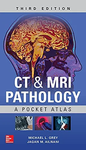
CT & MRI Pathology: A Pocket Atlas, 3rd Edition pairs cross-sectional images with concise descriptions of disease processes. Michael L. Grey and Jagan Mohan Ailinani organize pathology by body system, covering characteristic findings and potential mimics. Radiology learners, residents, technologists, and clinicians can use the image-focused format to study imaging-pathology relationships.
Inside this title
CT and MRI examples organized by anatomical body system
Current listed price $24.00Regular price $49.00Digital ebook
This atlas connects appearances on CT and MRI with the conditions that produce them. Images organized by body system accompany short accounts of pathology, with arrows and labels directing attention to relevant findings. The emphasis on visual examples supports comparison between disease patterns and normal variants that can complicate interpretation.
Coverage includes inflammatory, neoplastic, vascular, degenerative, and traumatic conditions, with discussion of signal patterns and contrast enhancement. The third edition's compact reference structure suits focused review of a region or condition. Radiology residents, medical students, technologists, and clinicians studying cross-sectional imaging can use the atlas to relate image characteristics to underlying disease processes.
Study guide included
Inside CT & MRI Pathology: A Pocket Atlas, 3rd Edition.
Grey and Ailinani teach image reading through paired pictures and short explanations, arranged by body region. The atlas insists on a sequence: establish modality, sequence and plane, describe location and signal, then compare the observation with entries that resemble it before anyone commits to a diagnosis.
Two opening parts explain what creates contrast in CT and in MRI, and how iodinated and gadolinium agents change what a reader sees. Regional parts then work through the central nervous system, head and neck, chest and mediastinum, abdomen, pelvis, and musculoskeletal structures. Each short entry couples images with a description, typical presentation, differential and, where useful, treatment and outlook. The final part steps back from individual entries to describe a repeatable method: state the technical parameters, describe the finding in neutral language, then weigh alternatives. Description is the durable skill; recognition follows from it.
What produces the signal
The first parts establish that brightness has no fixed meaning. CT reconstructs differences in x-ray attenuation, which favors dense structures, bone detail and several acute findings. MRI derives signal from tissue behavior in a magnetic field and changes contrast with pulse sequence. A bright area on a T1-weighted image and a bright area on a T2-weighted image are different observations. Contrast agents alter appearance by distinct mechanisms and carry their own safety considerations.
Name modality and sequence before interpreting
Density and signal are not synonyms
Ask what contrast was given and when
Parts I–II · PDF pp. 21, 25
Brain and spine by site
The nervous system section groups findings as tumor, vascular disease, congenital change, infection and degeneration, but anatomy comes first. Beginning with the compartment and neighboring structures makes displacement, edema and extension describable before a label is chosen. A lesion near the internal auditory canal raises questions unlike those raised by a white-matter focus. Spinal entries require several planes to separate bone, disc, soft tissue and cord.
Compartment narrows the differential quickly
Describe effects on adjacent structures
Use a second plane to place a finding
Part III · PDF p. 32
Head, neck and chest spaces
Head and neck entries span congenital lesions, inflammation and tumors, and the useful first step is naming the space involved and its relation to air, bone, glands and fat. A parotid mass and a sinus process can look broadly similar while differing in origin. The chest material separates lung, pleural, mediastinal, cardiovascular and breast findings, and reformatted vascular images help show extent and branch relationships. Value depends on the clinical question asked.
Identify the space before naming a process
Sketch the boundary between two examples
Match the technique to the question
Parts IV–V · PDF pp. 185, 215
Abdominal organs and phases
Abdominal coverage moves through bowel, liver, biliary tract, pancreas, kidneys, adrenal glands and soft tissue. The first question is origin: organ, duct, wall of a hollow structure or surrounding fat. Noting effects on adjacent structures supports comparison across inflammatory, obstructive, traumatic and neoplastic examples. Contrast phase matters too, since the same lesion can look different as enhancement evolves, and MR cholangiopancreatography answers a different question by highlighting fluid-filled ducts.
Establish the organ of origin first
Watch enhancement change across phases
Describe the pattern before the diagnosis
Part VI · PDF p. 279
Pelvis, joints and soft tissue
Pelvic entries are grouped by gynecologic, urologic and related pathology, and interpretation depends on identifying the organ and its boundaries. Localized disease and extension into neighboring structures call for different descriptions. Musculoskeletal entries range from fracture and degeneration to tendon injury, infection and tumor. CT shows bone geometry well, while MRI reveals marrow, ligament and tendon detail, so an organized review of bone, ligament, tendon and fluid pays off.
Locate the organ before characterizing the lesion
Choose CT or MRI by tissue of interest
Review bone, ligament, tendon and fluid
Parts VII–VIII · PDF pp. 407, 431
A method for reading images
The closing part converts the atlas into a procedure. State modality, plane, region and sequence or phase; then describe location, shape, attenuation or signal, enhancement and effects on neighbors. Compare that description with regional entries that look similar, and name the distinguishing feature. Because each entry ties appearance to presentation, cause, differential and outlook, the same visual pattern can carry different weight. The method also asks what the history leaves unresolved.
Describe before you compare
Name the feature that separates two options
Say what additional information would change it
Regional entries · Parts III–VIII
Chapter and section map
Principles of Imaging in CT and MRIPart I
Contrast MediaPart II
Central Nervous SystemPart III
Head and NeckPart IV
Chest and MediastinumPart V
AbdomenPart VI
PelvisPart VII
MusculoskeletalPart VIII
Key takeaways
Establish modality, sequence, plane and contrast phase before giving brightness any meaning.
Let anatomical compartment cut the list of plausible comparisons down to a manageable few.
Keep the visible feature separate from the inference, and state what the study cannot settle.
Radiology residents and technologists, medical students on imaging rotations, and clinicians who want a compact reference for matching cross-sectional appearances to common pathology at the scanner.
Topics covered
CT imaging
MRI sequences
cross-sectional anatomy
radiology atlas
contrast media
neuroradiology basics
musculoskeletal imaging
differential diagnosis
Questions about this ebook.
Edition, audience, and study-guide details at a glance.
What is CT & MRI Pathology: A Pocket Atlas, 3rd Edition about?
CT & MRI Pathology: A Pocket Atlas, 3rd Edition pairs cross-sectional images with concise descriptions of disease processes. Michael L. Grey and Jagan Mohan Ailinani organize pathology by body system, covering characteristic findings and potential mimics. Radiology learners, residents, technologists, and clinicians can use the image-focused format to study imaging-pathology relationships.
Which edition of CT & MRI Pathology: A Pocket Atlas, 3rd Edition is this?
This listing is for the 3rd edition. Check the title, ISBN, and book details above before ordering, especially if your course or reading list names a specific edition.
Who is CT & MRI Pathology: A Pocket Atlas, 3rd Edition for?
Radiology residents and technologists, medical students on imaging rotations, and clinicians who want a compact reference for matching cross-sectional appearances to common pathology at the scanner.
Does this page include a study guide?
Yes. This page includes an original study guide with six thematic sections, key takeaways, and a chapter and section map for structured reading.